A team of WID and UW–Madison researchers has induced human embryonic stem cells (hESC) to differentiate toward pure-population, mature heart muscle cells, or cardiomyocytes.
A substrate patterned with a precisely sized series of channels played a critical role in the advance.
Published online in the journal Biomaterials, the research could open the door to advances in areas that include tissue engineering and drug discovery and testing.

Researchers currently can differentiate hESC into immature heart muscle cells. Those cells, however, don’t develop the robust internal structures — repeating sections of muscle cells called sarcomeres — that enable cardiomyocytes to produce the contracting force that allows the heart to pump blood. Other cell components that allow heart muscle cells to communicate and work together also are less developed in immature cardiomyocytes.
One barrier to efforts to produce more mature cells is the culture surface itself; hESCs are notoriously finicky.
“It’s really hard to culture stem cells effectively and to provide them with an environment that’s going to help them to thrive and differentiate in the way you want,” says lead author Wendy Crone, WID BIONATES collaborator, professor of engineering physics, biomedical engineering and materials science and engineering at UW-Madison.
Recently, three-dimensional and micropatterned substrates have emerged as more accurately mimicking the cells’ physiological environment. However, the majority of previous research studies using patterning were conducted using cells from rats, says Max Salick, a Ph.D. student in materials science at WID and first author on the paper.

“One of the unique aspects of our research is that it observes human cardiomyocytes’ response to micropatterning geometries,” he says.
Working in partnership with other WID researchers and scientists at the Morgridge Institute for Research, the team focused on finding the pattern, including the right size scale, that suits the human stem cells.
“Our hypothesis was that if we could control the cell shape and how they bind to their surroundings using this micropatterning, we could coax them into forming more aligned, structurally sound fibrous structures that are more relevant in the heart,” says Salick.
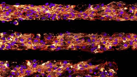
The researchers’ micropatterned substrate consists of a series of lanes, or channels. When they put the cells into the lanes, they saw a clear differences in how the cells responded to various lane sizes-and identifying the optimal size scale was key.
“If the lane was too wide, the cells weren’t really able to ‘feel’ their lane, so they didn’t align as well,” says Salick. “But with lanes less than 100 microns wide, we really started to see the alignment, a stronger sarcomere structure and a more mature phenotype.”
“It’s the closest we’ve gotten to pure-population adult cardiomyocytes.”
–Wendy Crone, BIONATES collaborator
The substrate method is more effective and easy to control than others the researchers have explored in the past. And now that they know lane width is critical, the researchers can make the lanes infinitely long, which enables individual cells to link and communicate with neighboring cells.
“This not only gets them to look like sarcomeres, and their internal structure starts to look like what it’s supposed to and behave like what it’s supposed to, but the cells also communicate with their neighbors,” says Crone. “It’s the closest we’ve gotten to pure-population adult cardiomyocytes.”
The National Institutes of Health and the UW–Madison Graduate School provided funding for the research. Collaborators on the project included Randolph Ashton, WID researcher and assistant professor of biomedical engineering; Timothy Kamp, a cardiology professor; students Brett N. Napiwocki, Gavin T. Knight and Shahzad A. Chindhy; and recent graduate Jin Sha.
—Renee Meiller, UW–Madison College of Engineering
You must be logged in to post a comment.