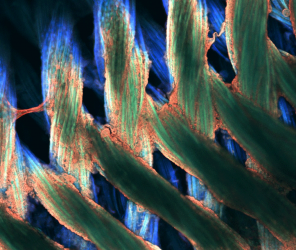
As a graduate student working with Professor Wendy Crone at WID, Max Salick used pluripotent stem cells to create human heart cells and explore their structure and integration with healthy hearts. In 2014, Max’s image of nano-fibers marked with a fluorescent stain was a winner in the Cool Science Image Contest at UW-Madison. After graduating in the fall of 2014, Max took a position as a postdoctoral researcher in the Neuroscience Department at the Novartis Institutes for Biomedical Research in Cambridge, MA. Under the guidance of Ajamete Kaykas and Ricardo Dolmetsch of the Neuroscience group, he has kicked off a project to model human neurological diseases using three-dimensional cerebral organoid tissues. As a result, the diseases that he wishes to replicate will more accurately mimic the phenotype that naturally occurs within native tissues.
What did you work on at WID?
Through several collaborations and with the support of my colleagues at WID, I was able to make interesting findings regarding cytoskeletal assembly. Cytoskeletal proteins allow a cell to control its shape and the forces that it produces, and I studied how their assembly changes in response to their surrounding mechanical environment. Using these findings, I found ways to more accurately mimic the cytoskeletal structure that naturally self-assembles within the healthy heart.
What are your tools for analysis?
With the incredible support I’ve received from UW-Madison, WID, and now from Novartis, I have been fortunate to have a massive array of tools at my disposal. There is a seemingly infinite complexity behind the biology of myocardial/neurological development and disease. Thus, these powerful tools are needed to answer the key questions regarding how cells interact with each other and their environment in both healthy and pathological conditions.
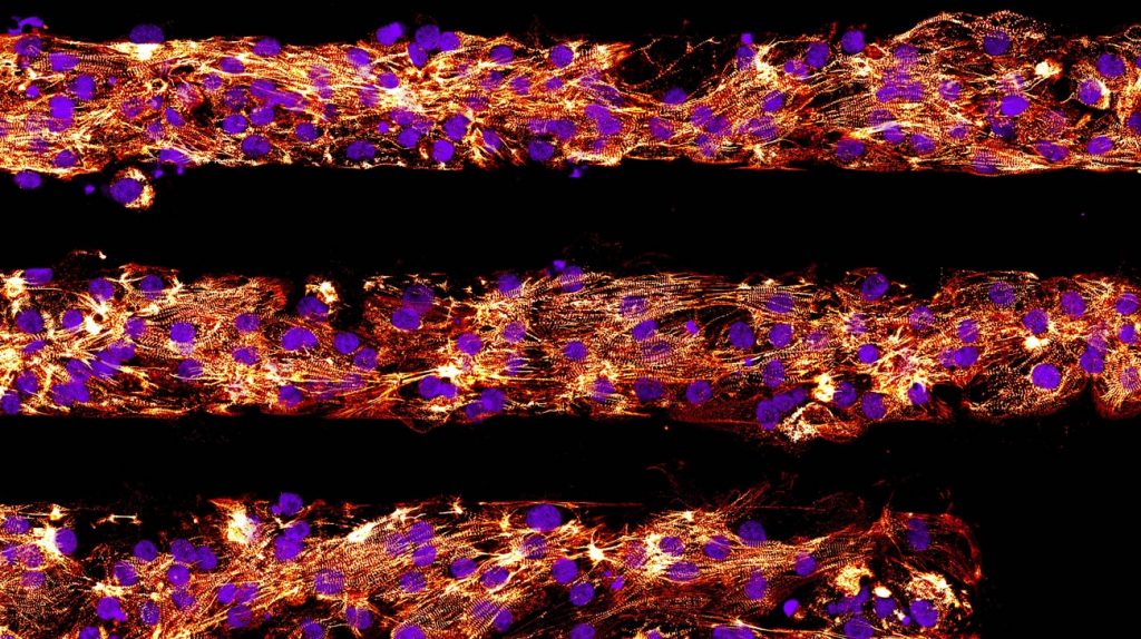
One of my favorite tools has always been microscopy; by fluorescently tagging proteins, we are able to see how they interact at the sub-micron scale, and the results can be as beautiful as they are informative. Additionally, with the emergence of more advanced microscopes, such as multi-photon, ultra-resolution, and lightsheet microscopes, I am now able to see even further, smaller, and faster than ever before.
“…it is always nice to get my hands dirty once in a while.”
-Max Salick
Another set of tools that I use has emerged recently from the field of genetics. Between advances in genome editing and next-generation sequencing technologies, we have been able to edit and analyze the genome in exciting, promising new ways. For this type of work, I utilize engineered e. coli as “DNA factories,” as well as several extremely advanced purifying and analyzing machines that allow us to examine the genome at single-nucleotide precision. At Novartis, I’ve been utilizing CRISPR/Cas9 technologies to conduct such genome editing, which allows me to make customized cell lines that exhibit useful genetic traits. For example, I can label specific, disease-linked proteins with fluorophores or create knock-out cell lines that exhibit mutations that have been discovered to contribute to neurological disease. In a way, these cell lines thus become the tools that I use for further analysis and experimentation.
When it is time to quantify my results and draw conclusions, I typically rely on image analysis software such as ImageJ, CellProfiler, and FIJI, as well as MATLAB for more computation-heavy work.
Lastly, when I find it necessary to construct custom equipment or to replace a particularly expensive component that I clumsily dropped, I am always eager to spend some quality time on SolidWorks and in the machine shop. After spending countless hours in extremely clean lab environments, it is always nice to get my hands dirty once in a while.
Tools for Writing?
Seclusion and caffeine. My first step in starting any writing project, whether it’s a publication or a grant proposal, is to get out of the lab. I turn my phone off and find a distraction-free corner somewhere in a library or coffee shop. That’s where I can break my writer’s block and, as a result, that’s where I can get my thoughts down on paper. I use Microsoft Word for writing and formatting, Mendeley for citation management, and PowerPoint to get images and scalebars properly arranged.
Tools for Collaboration?
I have always loved the collaborative atmosphere of UW-Madison and WID, and have been pleasantly surprised that this same level of collaboration occurs within industry at Novartis, as well. Some of the greatest tools for collaboration come from one’s personality. In a research setting, one is constantly surrounded by experts at their craft, and with a friendly and outgoing attitude, it is easy to form excellent relationships with such people that benefit both sides.
I would also consider the scientific communities as collaborative tools. For example, I happily ask and answer questions on forums such as Stack Overflow and ResearchGate. There are other tools that are shared openly, such as CRISPR Design tools provided by the Zhang Lab of MIT, human genome browsers provided by UC-Santa Cruz, the Brain Atlas provided by the Allen Institute, and a wide selection of image analysis scripts provided by groups such as LOCI at UW-Madison. Rather than forcing each group to develop these services on their own, these collaborations allow researchers to work together toward the common goal of ending human suffering more rapidly and efficiently.
Your ultimate tool for discovery?
I believe that tenacity, enthusiasm, and genuine curiosity are prerequisites for a scientist, but it is ultimately compassion that drives me. Every day, I consider the patients and their families that we are essentially trying to help. Even if only for a few seconds, it is this thought that drives me and makes me eager to get in to the lab every morning, thrilled to see what the day’s experiments will reveal.
— curated by Nolan Lendved
You must be logged in to post a comment.